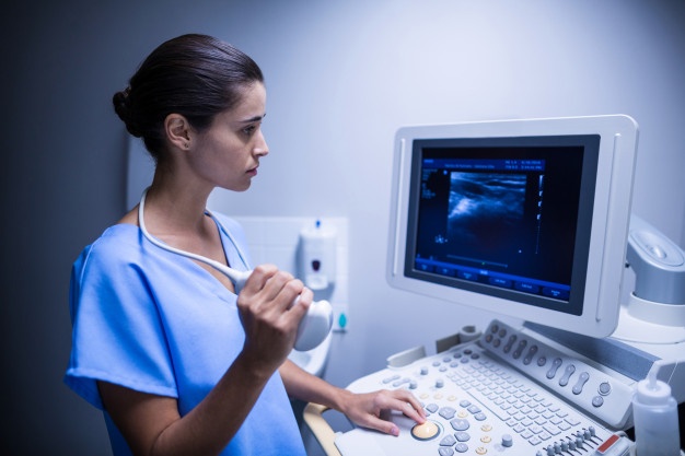
Radiology
Radiology is the field of medicine that makes use of magnetic waves, X-rays, and ultrasound to get clear images of the interior part of our body. Doctors then can use these images to diagnose ailments and illnesses as well as aid in developing treatment strategies.
Radiology facilities available at Ruby Medical Services
- X Ray : Chest, Spine, Head etc
- UltraSonography (USG)
- Mammography
- 2D Echo
- Colour Doppler
- CT Scan
- MRI
Computed Tomography (CT) Scan
CT is a computerized tomography (CT) can be described as an imaging test that generates precise pictures of bones, internal organs, and tissues. The images created during the CT scan are able to be transformed into three-dimensional images that give utmost clarity to treating Doctors to understand the problem with pin point accuracy and render pinpoint accurate treatment
Mammography
Mammography is an instrument to identify breast cancer or other anomalies The images reviewed by a radiologist are clearer and sharper than film x-rays , piece.
Magnetic Resonance Imaging (MRI)
Magnetic resonance imaging utilizes radio waves and a powerful magnetic field to provide crisp, precise images of organs and tissues inside. Since X-rays do not use and radiation exposure is not required. Instead radio waves hit toward the body. The test takes between 30 and 50 minutes in the average, and comprises of a variety of imaging studies. A majority of studies require a tiny intravenous injection of the contrast agent. But, this contrast agent is not iodine-based which is a component that is used in other contrast agents for Xrays or CT scans. MRI is commonly used to detect or assess cancers and other diseases of the liver, the heart and bowel. It can also be employed to monitor the development of a baby still during the womb.
X-ray (Radiography)
X-ray (also known as radiography) utilizes a tiny amount of radiation to create images of the inside of your body. These are the most widely used method of medical imaging and are also the most ancient. They are commonly employed to detect bone fractures, injuries or infections. They can also be used to detect foreign bodies in soft tissues. In certain instances the x-ray test is used along with a contrast material based on iodine that is swallowed to assist doctors in seeing specific organs blood veins or tissues.
SONOGRAPHY AND COLOUR DOPPLER
The Sonography also known as an Ultrasound examination consists of high-frequency sound waves that take images of inside your body. Sonographies are secure, affordable and don’t present to your body for radiation. This provides an accurate image, which leads to an accurate diagnosis. Apart from high-resolution Ultrasounds We also provide Doppler scans in routine Ultrasound examinations, aswell in peripheral arterial and venous Doppler studies as well as the carotid artery Doppler as well as cardiac Doppler scans.


